
Not every painful knee in a 55-year-old is arthritis. A subchondral insufficiency fracture of the knee (SIFK) is a stress injury of the bone just beneath the cartilage, most often on the medial femoral condyle, and it tends to announce itself as sudden medial knee pain with no clear injury, present with walking and often at rest. Plain radiographs can look nearly normal while a T2 MRI shows a bright zone of marrow edema and a dark subchondral fracture line. For years this picture was called spontaneous osteonecrosis of the knee, or SONK. Pathology and MRI studies have since shown that the bone is not dying first; it is fracturing under load, and the necrosis seen in advanced cases is a consequence rather than the cause.
The distinction matters because a fracture can heal if it is protected and supported, while a collapsed condyle generally cannot. In the video below, Dr. Jeremy Burnham, an orthopedic sports medicine surgeon at Ochsner-Andrews Sports Medicine Institute in Baton Rouge, Louisiana, presents a 58-year-old woman whose medial femoral condyle SIFK did not settle with protected weight bearing, a home exercise program, and bone health treatment, and demonstrates an arthroscopically assisted, percutaneous fixation technique using bio-integrative OSSIOfiber trimmable fixation nails placed across the lesion. This article explains what the condition is, what the natural history data show, where this technique fits among the options, and what the evidence does and does not yet support.
📋 In This Article
- What Is a Subchondral Insufficiency Fracture of the Knee?
- Watch: Arthroscopically Assisted Percutaneous Fixation (Surgical Technique Video)
- Why SIFK Matters: Natural History and Risk of Collapse
- How MRI Defines the Lesion and Guides Treatment
- Nonoperative Treatment Comes First
- Why Support the Subchondral Bone? The Rationale for Bio-Integrative Fixation
- The Case: A 58-Year-Old With Persistent Medial Knee Pain
- How Dr. Burnham Performs the Procedure
- What the Evidence Does and Does Not Show
- Potential Disadvantages and Who Is Not a Candidate
- The Bottom Line
- References
- Frequently Asked Questions
What Is a Subchondral Insufficiency Fracture of the Knee?
Subchondral bone is the thin plate of bone directly under the articular cartilage. It absorbs and distributes the load that passes through the joint with every step. When repetitive load exceeds what that bone can tolerate, often because the meniscus that normally cushions the compartment is torn or extruded, the subchondral plate can crack. The result is an insufficiency fracture: a fracture of weakened or overloaded bone under normal activity, rather than a fracture from a fall or collision.
A 2022 review in the Japanese Journal of Radiology traces the terminology. The condition was named spontaneous osteonecrosis of the knee in the era before MRI, when surgeons saw a collapsed, dead segment of condyle and reasonably assumed a vascular cause. MRI changed the picture: the earliest finding is a subchondral T2 hypointense fracture line surrounded by extensive bone marrow edema, and SONK is now understood as the advanced, collapsed end of the SIFK spectrum (Ochi et al., Jpn J Radiol 2022). The same review notes that chondral lesions and meniscal abnormalities, particularly posterior root tears, are found in many patients with SIFK and are thought to contribute to its development. For a patient, the practical translation is that a meniscus problem and a bone problem often arrive together, and the bone problem is the one that threatens the joint. Dr. Burnham’s meniscus tear guide covers the root tear side of that equation.
Watch: Arthroscopically Assisted Percutaneous Fixation (Surgical Technique Video)
The video runs just over nine minutes. The first two and a half minutes cover the condition, the nonoperative pathway, and the rationale and trade-offs of fixation. The case presentation and imaging follow, then the surgical technique step by step, and the video closes with pearls, pitfalls, and the postoperative plan. Surgical footage is shown.

Chapters in the video: what a subchondral insufficiency fracture is (0:32), nonoperative treatment first (1:02), rationale for bio-integrative fixation (1:13), advantages and disadvantages (1:44 to 2:32), the case and imaging (2:32), the surgical sequence (3:29 to 6:55), pearls and pitfalls (6:55), final imaging (7:42), and the postoperative plan (7:58).
Why SIFK Matters: Natural History and Risk of Collapse
The reason SIFK deserves attention is what happens when it is left to run its course. The largest natural history series comes from the Mayo Clinic, which reviewed 223 patients with an MRI-confirmed diagnosis, mean age 65, 71% female. The medial compartment was involved in 89% of knees, and 74% of medial menisci had a root or radial tear. Over a mean of about three years, 34% of patients progressed to surgery and 30% underwent knee arthroplasty; among those followed more than five years, those figures rose to 47% and 37%. Baseline arthritis, older age, fractures on both the femoral condyle and the tibial plateau in the same compartment, meniscal extrusion, and varus malalignment were each associated with progression to arthroplasty (Pareek et al., J Bone Joint Surg Am 2020).
Not every lesion behaves this way. In a 50-patient MRI series, 78% of SIFK lesions were low grade and 70% were in the medial femoral condyle. High-grade lesions were marked by medial meniscus posterior root tears with moderate to severe extrusion, high-grade cartilage wear, larger lesion dimensions, and collapse of the articular surface, while improvement of marrow edema on a follow-up MRI was highly predictive of low-grade disease (Sayyid et al., Skeletal Radiol 2019). A later 219-patient study confirmed that high-grade SIFK travels with high-grade cartilage wear in the same compartment (Allam et al., Skeletal Radiol 2021). The window for a joint-preserving option is therefore early, before the subchondral plate collapses and before the cartilage above it is lost.
How MRI Defines the Lesion and Guides Treatment
Radiographs come first, mainly to measure alignment, look for joint space loss, and exclude a collapsed condyle or advanced arthritis. In the case shown, standing views showed preserved alignment and no substantial joint space narrowing that could explain the severity and focal location of the symptoms. That mismatch between a nearly normal radiograph and a patient who cannot bear weight comfortably is the classic prompt to obtain an MRI.

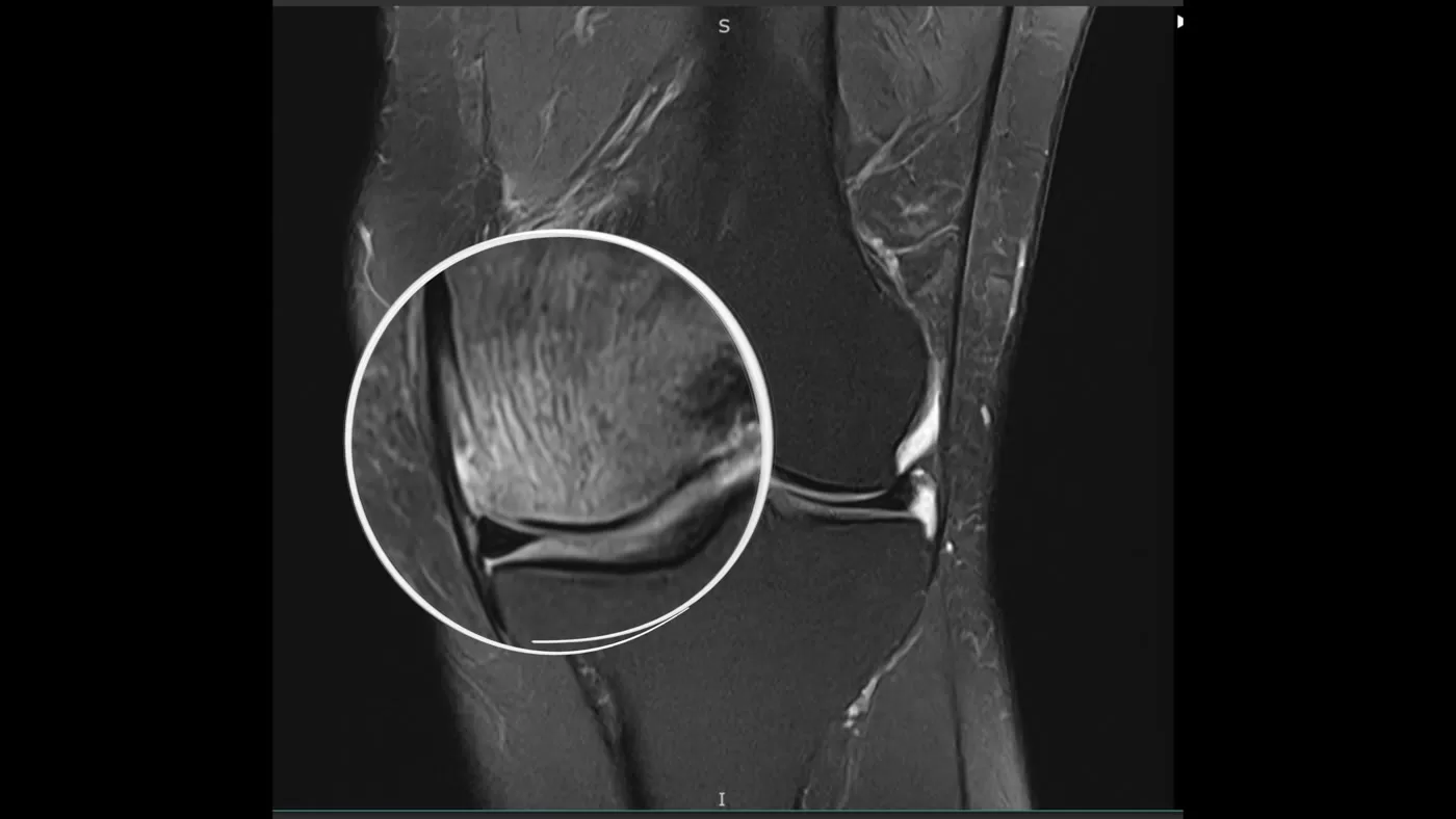
On fluid-sensitive (T2 or STIR) sequences, SIFK appears as a focal zone of bright marrow edema beneath the articular surface with a dark subchondral line that represents the fracture itself. Each plane answers a different question. The coronal plane shows how far the lesion extends from medial to lateral. The sagittal plane shows its front-to-back extent and its relationship to the weight-bearing surface. The axial plane completes the three-dimensional picture. When fixation is planned, those three planes are what determine how many implants are needed and the angle of each trajectory. The MRI also documents the meniscus and the cartilage, which, as the natural history data above show, carry much of the prognosis.
Nonoperative Treatment Comes First
Because a subchondral insufficiency fracture is a fracture, the first-line treatment is the same principle applied to any stress fracture: take the load off and give the bone a chance to heal. Dr. Burnham’s practice begins with protected weight bearing on crutches, activity modification, physical therapy and a home exercise program, bracing when appropriate, and attention to overall bone health, including evaluation and treatment of low bone density where indicated. Many lower-grade lesions improve on this plan, and a follow-up MRI showing resolving marrow edema is a reassuring sign that the lesion is healing rather than progressing (Sayyid et al., Skeletal Radiol 2019).
Surgery enters the conversation when pain persists despite that program, the joint surface remains intact, and the knee is otherwise worth preserving: alignment is acceptable, arthritis is limited, and the patient is motivated to avoid or defer a knee replacement. In the matched cohorts reported by the Mayo group, patients with SIFK treated nonoperatively had a 2-year arthroplasty-free survival of 61%, and only 15% among those with high-risk SIFK scores (Pareek et al., J Knee Surg 2023). Those numbers frame the decision for a patient in her fifties who wants to keep her own knee.
Why Support the Subchondral Bone? The Rationale for Bio-Integrative Fixation
The most studied surgical option for a painful subchondral lesion has been subchondroplasty, in which a calcium phosphate bone substitute is injected into the lesion under fluoroscopy to stiffen the bone while it remodels. A systematic review of 17 studies and 756 patients found that pain scores fell from a mean of 7.8 to 3.4 on a 10-point scale and that function improved, but nearly all studies were low-level evidence and conversion to knee arthroplasty ranged from 12.5% to 30% across follow-up periods of 10 months to 7 years (Nairn et al., KSSTA 2021). In a 216-patient single-surgeon series, survivorship free of arthroplasty was 73% at five years overall, falling to 62% in patients with grade 4 cartilage lesions and 59% in those over 50 with grade 4 lesions (Wood et al., Clin Orthop Relat Res 2023). A prospective multicenter cohort of 70 patients reported 76.2% freedom from arthroplasty at two years (Cohen et al., J Knee Surg 2025).
The technique shown here takes a different mechanical approach. Rather than filling the lesion, it bridges it. Rigid implants are placed percutaneously across the injured subchondral bone, from the medial cortex of the condyle to the cortex at the edge of the intercondylar notch, in a rafter-like arrangement beneath the articular surface. The goal is to share load across the weakened plate, the way a beam supports a sagging floor, so the fracture can heal under protected weight bearing without collapsing. The implants are OSSIOfiber trimmable fixation nails, a fiber-reinforced implant built from a matrix of continuous mineral fibers that is designed to be gradually replaced by the patient’s own bone rather than remaining as permanent metal. The manufacturer markets this application as the KneeBar procedure. The fixation nail family received FDA 510(k) clearance in 2019 for maintenance of alignment and fixation of bone fractures, osteotomies, arthrodeses, and bone grafts with appropriate additional immobilization (OSSIO, July 2020), and its use for a subchondral fracture falls within that fracture-fixation indication.
Two features make the implant attractive for this location. First, because it is radiolucent and does not generate the artifact of metal, follow-up MRI of the condyle remains readable. Second, because it is designed to integrate, there is no permanent hardware to remove or to complicate a later arthroplasty if one is eventually needed. The integration itself has been documented in humans at a different anatomic site: in 24 patients who received the same material for hammertoe fusion, the implant was no longer visible on imaging in 21% and only partially visible in 33% at two years, with the border between implant and bone no longer distinguishable in 88% and no cyst formation or adverse implant-related findings (Stalc et al., Foot Ankle Surg 2022). In a sheep ACL reconstruction model, mineral fiber screws showed tissue ingrowth by 28 weeks and were fully replaced by remodeled bone at 132 weeks without a pro-inflammatory response (Cole et al., Sci Rep 2024). The manufacturer describes complete integration within roughly 24 months; in the video Dr. Burnham frames it as approximately 18 to 24 months.
The Case: A 58-Year-Old With Persistent Medial Knee Pain
The patient in the video is a 58-year-old woman with approximately two months of atraumatic medial-sided left knee pain, present with walking and at rest. Examination localized tenderness to the medial femoral condyle and the medial joint line. Her pain persisted despite protected weight bearing with crutches, a home exercise program, and medical treatment of bone health.
Standing radiographs showed preserved alignment, no acute bony abnormality, and no substantial joint space loss. Coronal, sagittal, and axial T2 MRI localized a subchondral insufficiency fracture to the medial femoral condyle and defined its full extent. Diagnostic arthroscopy at the time of surgery found intact cruciate ligaments, no unstable meniscal tear requiring treatment, and grade 2 cartilage wear in the weight-bearing zone of the medial femoral condyle with grade 2 changes in the lateral and patellofemoral compartments. A preserved joint surface over the lesion, acceptable alignment, and only moderate cartilage wear are what made her a candidate for a joint-preserving procedure rather than a replacement.
How Dr. Burnham Performs the Procedure
The construct is planned in three dimensions from the MRI and executed with intraoperative fluoroscopy and arthroscopy working together. Every trajectory has to satisfy three rules: stay within the subchondral bone, protect the articular surface, and end safely at the cortex bordering the intercondylar notch. The steps shown in the video are:
- Diagnostic arthroscopy. The joint is assessed systematically. Unstable cartilage flaps are debrided to stable margins while viable cartilage is preserved, and hypertrophic synovium is addressed as needed.
- Spinal needle localization. A spinal needle translates the MRI target to the arthroscopic surface and the fluoroscopic position, confirming the percutaneous entry point and trajectory before any pin is placed.
- Guide pin placement. Starting anteriorly, a guide pin is advanced across the medial femoral condyle toward the notch-side cortex. Orthogonal fluoroscopic views confirm that the pin sits beneath the articular surface at the intended depth and does not penetrate the notch. Both planes are confirmed before drilling.
- Drilling and measuring. With the pin held in place, a cannulated drill prepares the channel to the notch-side cortex. The measuring guide is seated directly against the femoral cortex so the implant length spans the condyle and reaches the intended cortical endpoint.
- Trimming the implant. The trimmable fixation nail is cut to the measured length, and the leading end can be trimmed to ease insertion. Measurement and cut length are double-checked before delivery.
- Seating the implant. The implant is advanced over the guide pin under fluoroscopic and arthroscopic visualization. The near end is seated flush at the medial condyle and the far end engages the cortex bordering the notch without penetrating the notch surface.
- Repeat for three implants. The sequence is repeated twice more, with trajectories progressing posteriorly to span the zone of injury while maintaining safe separation. Each implant is planned and verified independently.
- Final verification. Orthogonal fluoroscopy confirms all three implants and their cortical endpoints, and repeat arthroscopy confirms that nothing is prominent in the condyle or the notch and that no debris remains in the joint.
Dr. Burnham’s pearls for the procedure, from the video: plan the construct in all three MRI planes; use the spinal needle to translate the lesion to the operative field; confirm every guide pin on orthogonal views before drilling and keep the pin in place through channel preparation and measurement; seat the measuring guide directly on the cortex to avoid a wrong implant length; protect both the articular surface and the notch; keep safe separation between trajectories; and expect subchondral bone quality to vary across the condyle, relying on tactile feedback together with imaging rather than assuming every channel will feel the same. The common pitfalls are the mirror image of those pearls: assuming each trajectory will behave identically, losing the established guide pin position, and advancing an implant without independent fluoroscopic and arthroscopic confirmation.
Postoperatively, the patient was foot-flat weight bearing for two weeks and then advanced as tolerated, with range of motion as tolerated from the start. Physical therapy began on postoperative day two or three, aspirin was used for blood clot prophylaxis, and the first follow-up visit was at 10 to 14 days.
What the Evidence Does and Does Not Show
Patients and referring physicians deserve a plain statement of where this technique sits. The natural history of SIFK is well characterized, and the case for intervening before collapse in a joint that is otherwise worth saving rests on that data. The mechanical rationale for supporting the subchondral plate is sound, and the integration behavior of the implant material has been documented in humans at other sites and in large-animal models. What does not yet exist is a peer-reviewed clinical outcome series for OSSIOfiber fixation of subchondral insufficiency fractures of the knee. As of November 2026, the only registered study is a 10-patient observational study at Stanford and Rush, which began enrolling in October 2025 with primary completion expected in mid-2027 (ClinicalTrials.gov NCT06138548). The manufacturer cites a pilot study showing improved pain and function at 6 and 12 months, but that work is reported as data on file and in press rather than as published, peer-reviewed results.
That is why Dr. Burnham states in the video that long-term clinical evidence remains limited, and why the procedure is offered as an individualized option for selected patients rather than as a standard of care. The closest comparator with published outcomes, subchondroplasty, shows meaningful pain relief in most patients but a conversion to arthroplasty of roughly one in four by five years, with worse results in patients over 50 who already have full-thickness cartilage loss (Wood et al., CORR 2023). Those are the benchmarks any subchondral procedure, including this one, will need to meet or beat as data accumulate.
Potential Disadvantages and Who Is Not a Candidate
The procedure carries the risks inherent to any knee surgery and to fluoroscopically guided drilling, including implant malposition and the possibility of breaching the articular surface or the intercondylar notch, which the technique above is designed to prevent. Protected weight bearing and a rehabilitation program are still required, and healing of the fracture and relief of symptoms are not guaranteed. Because the implant is designed to integrate over many months, the bone must be protected while that happens.
Equally important is who should not be offered it. Subchondral fixation is not intended to replace knee arthroplasty in a patient with advanced subchondral collapse, substantial or diffuse arthritis, or major malalignment. Varus malalignment in particular was an independent predictor of progression to arthroplasty in the Mayo series (Pareek et al., JBJS 2020), and a knee that is overloading its medial compartment because of alignment needs that problem addressed, whether by realignment osteotomy or by replacement, rather than by supporting the bone alone. Patients with a large, unstable cartilage defect over the lesion are better served by cartilage restoration options or arthroplasty, depending on age and extent. For the right patient, early, with an intact surface and a preserved joint, fixation offers a chance to heal the bone and keep the knee.
About the Author
Jeremy M. Burnham, MD is a board-certified orthopedic surgeon and Director of Sports Medicine at Ochsner-Andrews Sports Medicine Institute in Baton Rouge, Louisiana, with a clinical focus on complex knee problems, joint preservation, and ACL reconstruction. Following his orthopedic surgery residency at the University of Kentucky, he completed his sports medicine fellowship at the University of Pittsburgh Medical Center (UPMC), where he trained under the late Dr. Freddie Fu, a pioneer of anatomic ACL reconstruction, Dr. James Bradley, a renowned sports medicine surgeon and longtime professional team orthopedist, and Dr. Volker Musahl, an internationally recognized ACL surgeon and researcher. His team physician experience spans professional sports teams, the University of Pittsburgh, and Southern University. With more than 145 peer-reviewed publications, book chapters, and scientific presentations, Dr. Burnham is the most published ACL surgeon in Louisiana. His research focuses on advancing ACL reconstruction, optimizing return-to-sport outcomes, and pioneering injury prevention, and has been recognized with the Game Changer Award from the Arthritis Foundation and the Playmaker Award from AOSSM. He serves as a site principal investigator for two federally funded clinical trials (NIH STABILITY 2 and Department of Defense STaR Trial). View full credentials and publications.
The Bottom Line
A subchondral insufficiency fracture of the knee is a stress fracture of the bone beneath the cartilage, not a dying bone, and that reframing is what opens the door to healing it. Left alone, roughly a third of patients progress to knee replacement within a few years, and the odds are worse with meniscal root tears, extrusion, varus alignment, and existing cartilage wear. Protected weight bearing, therapy, and bone health optimization remain the first line, and many lower-grade lesions settle with that plan.
For the patient whose pain persists while the joint surface is still intact, arthroscopically assisted percutaneous fixation with bio-integrative implants is a minimally invasive, joint-preserving option that supports the fracture while it heals and leaves no permanent hardware behind. The mechanical rationale is sound and the material’s integration is documented, but knee-specific clinical outcomes are still being collected, and the procedure is not a substitute for arthroplasty in advanced collapse, diffuse arthritis, or significant malalignment. Patients in Baton Rouge and across Louisiana with sudden, unexplained medial knee pain and a near-normal radiograph should ask for an MRI before accepting an arthritis label, because the diagnosis changes the options. Dr. Burnham’s guide to finding the right orthopedic doctor for knee pain covers the questions worth asking at that visit.
References
- Ochi J, Nozaki T, Nimura A, Yamaguchi T, Kitamura N. Subchondral insufficiency fracture of the knee: review of current concepts and radiological differential diagnoses. Jpn J Radiol. 2022;40(5):443-457. PubMed
- Pareek A, Parkes CW, Bernard C, Camp CL, Saris DBF, Stuart MJ, Krych AJ. Spontaneous Osteonecrosis/Subchondral Insufficiency Fractures of the Knee: High Rates of Conversion to Surgical Treatment and Arthroplasty. J Bone Joint Surg Am. 2020;102(9):821-829. PubMed
- Sayyid S, Younan Y, Sharma G, Singer A, Morrison W, Zoga A, Gonzalez FM. Subchondral insufficiency fracture of the knee: grading, risk factors, and outcome. Skeletal Radiol. 2019;48(12):1961-1974. PubMed
- Allam E, Boychev G, Aiyedipe S, Morrison W, Roedl JB, Singer AD, Gonzalez FM. Subchondral insufficiency fracture of the knee: unicompartmental correlation to meniscal pathology and degree of chondrosis by MRI. Skeletal Radiol. 2021;50(11):2185-2194. PubMed
- Pareek A, Parkes CW, Slynarski K, Walawski J, Smigielski R, van der Merwe W, Krych AJ. Risk of Arthroplasty in Patients with Subchondral Insufficiency Fractures of the Knee: A Matched Study of the Implantable Shock Absorber using a Validated Predictive Model. J Knee Surg. 2023;37(1):73-78. PubMed
- Nairn LN, Subramaniam M, Ekhtiari S, Axelrod DE, Grant JA, Khan M. Safety and early results of Subchondroplasty for the treatment of bone marrow lesions in osteoarthritis: a systematic review. Knee Surg Sports Traumatol Arthrosc. 2021;29(11):3599-3607. PubMed
- Wood DS, Paulson S, Nolan JR, Spanyer J, Harm RG, Heis FT. What Factors are Associated With Conversion to Knee Arthroplasty After Subchondroplasty? Clin Orthop Relat Res. 2023;481(8):1543-1550. PubMed
- Cohen SB, Hajnik C, Loren GJ, et al. Prospective Evaluation of Clinical Outcomes of the Subchondroplasty Procedure for Treatment of Symptomatic Bone Marrow Lesions of the Knee. J Knee Surg. 2025;38(6):290-299. PubMed
- Stalc J, Cicchinelli LD, Miller S, Sofka CM, Richter M. Fiber-reinforced fixation implant for proximal interphalangeal joint arthrodesis shows advanced implant bio-integration at 2-year follow-up. Foot Ankle Surg. 2022;28(8):1293-1299. PubMed
- Cole BJ, Easley JT, Nyska A, Rousselle S. Anterior cruciate ligament reconstruction in a translational model in sheep using biointegrative mineral fiber reinforced screws. Sci Rep. 2024;14(1):27408. PubMed
- Stanford University. Bio-Integrative, Fiber-Reinforced Kneebar for Treating Subchondral Insufficiency of the Knee. ClinicalTrials.gov identifier NCT06138548. Accessed November 8, 2026.
- OSSIO Ltd. OSSIO Announces U.S. Launch of Commercial OSSIOfiber Trimmable Fixation Nail. BusinessWire, July 22, 2020. Accessed November 8, 2026.
Frequently Asked Questions
What is a subchondral insufficiency fracture of the knee, and is it the same as SONK?
It is a stress fracture of the thin layer of bone directly beneath the knee cartilage, most often on the inner (medial) side of the thigh bone, usually in people over 50 and often alongside a torn or extruded meniscus. It was once called spontaneous osteonecrosis of the knee (SONK) because advanced cases showed dead, collapsed bone. MRI studies have shown that the fracture comes first and the bone damage follows, so SONK is now considered the late stage of the same condition.
Why does my X-ray look normal when my knee hurts so much?
Early subchondral insufficiency fractures are invisible on plain radiographs because the cartilage and the joint space are still intact. The injury shows up on MRI as a bright area of bone marrow swelling with a dark fracture line just under the joint surface. A near-normal X-ray in a patient with sudden, severe medial knee pain is exactly the situation in which Dr. Burnham orders an MRI, because an early diagnosis is what makes joint-preserving treatment possible.
Can a subchondral insufficiency fracture heal without surgery?
Many can, especially lower-grade lesions. First-line treatment is protected weight bearing on crutches, activity modification, physical therapy, bracing when appropriate, and attention to bone health. A follow-up MRI that shows the marrow swelling improving is a good sign the bone is healing. Surgery is considered when pain persists despite that program and the joint surface is still intact, because about a third of patients in the largest published series went on to knee replacement when the condition progressed.
What are the implants made of, and do they stay in my knee forever?
The implants are OSSIOfiber trimmable fixation nails, made from a matrix of continuous mineral fibers rather than metal. They are designed to support the bone while the fracture heals and then be gradually replaced by the patient’s own bone over roughly 18 to 24 months, so no permanent hardware remains. Because the material is not metal, follow-up MRI stays readable, and nothing needs to be removed if a knee replacement were ever needed later. Published two-year imaging in patients who received the material for toe surgery showed the implants progressively disappearing into bone.
Who is not a good candidate for this procedure?
Patients whose bone has already collapsed, who have significant or widespread arthritis, or who have major malalignment of the leg are not candidates, because supporting the bone alone will not fix those problems. In those situations Dr. Burnham discusses realignment osteotomy, cartilage restoration, or knee replacement instead. It is also important to understand that long-term clinical evidence for this specific technique is still limited, so it is offered as an individualized option for selected patients after nonoperative treatment has been tried.