
A full-thickness cartilage defect on the weight-bearing surface of the knee can hurt out of proportion to what plain radiographs show. The joint space looks preserved, the alignment is close to normal, and yet the patient has months of localized pain, swelling, and catching that physical therapy, aspirations, and injections have not settled. For a young, active patient, the treatment question is how to restore that one damaged area without burning bridges to the options that might be needed decades later.
In the video below, Dr. Jeremy Burnham, an orthopedic sports medicine surgeon at Ochsner-Andrews Sports Medicine Institute in Baton Rouge, Louisiana, walks through a single-stage cartilage restoration using two CartiHeal Agili-C implants to treat a 12 by 20 millimeter grade 4 lesion of the medial femoral condyle in a 28-year-old electrician. This article summarizes the evidence behind the implant, the patient-selection rules Dr. Burnham applies before offering it, and the technical details that determine whether a two-implant construct succeeds. Every clinical number links to its source.
📋 In This Article
- What Is the Agili-C Implant?
- Watch: Two-Implant Agili-C Cartilage Restoration (Surgical Video)
- Nonoperative Care Comes First
- Who Is a Candidate? Indications and Contraindications
- What the Randomized Trial Showed, and What It Did Not
- The Case: A 28-Year-Old Electrician
- How Dr. Burnham Performs a Two-Implant Agili-C Restoration
- Recovery and Early Follow-Up
- Limitations and Open Questions
- The Bottom Line
- References
- Frequently Asked Questions
What Is the Agili-C Implant?
Agili-C is a cell-free, biphasic scaffold made of aragonite, a crystalline form of calcium carbonate derived from coral. The upper phase sits at the level of the cartilage surface and the lower phase extends into the underlying bone, where its interconnected pores allow cells, blood vessels, and healing tissue from the surrounding bone to migrate in. The scaffold is biodegradable and is gradually resorbed as repair tissue develops. It is implanted in a single operation, press-fit into a prepared socket without screws, sutures, or permanent hardware, and it requires no cell harvest, no donor cartilage, and no second stage.
The U.S. Food and Drug Administration granted premarket approval to Agili-C on March 29, 2022 (PMA P210034) for patients 21 to 75 years old with International Cartilage Regeneration and Joint Preservation Society (ICRS) grade III or higher knee joint surface lesions totaling 1 to 7 square centimeters, in knees without severe osteoarthritis (Kellgren-Lawrence grade 0 to 3). A subchondral bone defect or cyst deeper than 8 millimeters is a contraindication. Agili-C is a product of CartiHeal, now part of Smith+Nephew.
Watch: Two-Implant Agili-C Cartilage Restoration (Surgical Video)
The video runs just under 11 minutes. The first four minutes cover how the implant works, who qualifies, and the imaging workup; the remainder is the operation itself, from diagnostic arthroscopy through closure, followed by the postoperative plan and 2-week radiographs. Surgical footage is shown.

Chapters in the video: how the scaffold works (0:50), indications and technical requirements (1:42), the case presentation (2:18), imaging (3:11), diagnostic arthroscopy (4:27), exposure and lesion sizing (4:47), planning two implant sites (5:15), preparing and filling the posterior site (5:23), the anterior site (7:06), final inspection (8:19), the postoperative plan and 2-week radiographs (9:06), and pearls (9:55).
Nonoperative Care Comes First
Cartilage restoration is a treatment for a specific problem, not for knee pain in general. Before surgery is discussed, Dr. Burnham’s team confirms that the symptoms actually match the lesion: pain and tenderness localized to the involved compartment, mechanical symptoms such as catching or swelling with activity, and a lesion on MRI in the location that explains them. Most patients have already completed a structured course of physical therapy, activity modification, and anti-inflammatory treatment, and many have had an aspiration or a corticosteroid injection. When those measures fail and the lesion remains the best explanation for recurring, compartment-specific symptoms, surgical restoration becomes a reasonable next step.
Three other factors are assessed before any implant is chosen, because a technically perfect repair can still be overloaded. Limb alignment is measured on standing long-leg radiographs; if the mechanical axis drives excessive force through the involved compartment, a realignment osteotomy may be needed alongside or instead of cartilage work (Dr. Burnham’s article on alignment and when osteotomy helps covers that decision). Ligament stability is confirmed on examination. And the menisci are evaluated, since a deficient meniscus shifts load onto the cartilage; Dr. Burnham’s meniscus tear guide explains why preservation matters.
Who Is a Candidate? Indications and Contraindications
The scaffold is not appropriate for every cartilage lesion. In Dr. Burnham’s practice, the lesion must be a contained grade 3 or 4 defect on the femoral condyle or trochlea with a complete wall of healthy bone at least 2 millimeters thick around each planned socket. A subchondral defect or cyst deeper than 8 millimeters rules the implant out, consistent with the FDA labeling. Because each socket must be prepared exactly perpendicular to the joint surface, the procedure is performed through a limited medial or lateral parapatellar mini-arthrotomy rather than purely arthroscopically. When a single lesion is too long for one implant, as in the case shown here, two scaffolds can be used, provided a bone bridge of at least 5 millimeters is preserved between them.
Alternatives exist for lesions that fall outside these criteria. Larger or uncontained defects, or those with deep bone loss, are often better served by osteochondral allograft transplantation, which replaces both cartilage and bone with donor tissue. Microfracture remains an option for small lesions, but its durability is the concern: a systematic analysis of 28 studies and 3,122 patients found reliable short-term improvement in the first 24 months with conflicting reports on whether that improvement lasts (Mithoefer et al., AJSM 2009).
What the Randomized Trial Showed, and What It Did Not
The pivotal evidence for Agili-C is a multicenter randomized controlled trial of 251 patients at 26 centers, published in the American Journal of Sports Medicine in 2023 (Altschuler et al., AJSM 2023). Patients aged 21 to 75 with up to three ICRS grade 3a or higher lesions on the femoral condyles or trochlea, a total treatable area of 1 to 7 square centimeters, bony defect depth of 8 millimeters or less, and Kellgren-Lawrence grade 0 to 3 were randomized 2 to 1 to the scaffold (167 patients) or to arthroscopic debridement or microfracture (84 patients). At 24 months:
- The implant group’s mean improvement in overall KOOS (Knee injury and Osteoarthritis Outcome Score), the primary endpoint, was about twice that of the control group, and the implant was superior on every secondary endpoint at every follow-up.
- Responder rate (at least a 30-point KOOS improvement) was 77.8% with the implant versus 33.6% with debridement or microfracture.
- Defect fill of at least 75% on MRI was seen in 88.5% of implant patients versus 30.9% of controls.
- Failure, defined as any secondary treatment, was 7.2% in the implant group versus 21.4% in controls.
Five-year results from the same trial were published in 2026 (Altschuler et al., AJSM 2026). Overall KOOS at final follow-up was 81.0 in the implant group versus 59.1 in the control group, a reported 22.6-point difference, with responder rates of 74.7% versus 29.6% and failure rates of 15.0% versus 35.7%. Among the subgroup with mild to moderate osteoarthritis, the implant group still had a higher responder rate (74.6% versus 36.2%) and a lower failure rate (13.2% versus 40.7%). An earlier prospective series of 86 patients with Kellgren-Lawrence grade 2 or 3 osteoarthritis reported KOOS Pain improving from 49.6 to 79.5 at two years, with 9.3% requiring revision (Kon et al., AJSM 2021), and a sex-based analysis of the trial at four years found women and men achieved comparable results (Kon et al., J Orthop Traumatol 2025).
Two points keep these results in perspective. First, the comparator was debridement or microfracture, not osteochondral allograft or autologous chondrocyte implantation, so the trial does not establish superiority over the other restorative options a surgeon might consider for the same lesion. Second, the trial excluded knees with severe arthritis and deep bony defects, so the outcomes apply to the patients who meet the indications above and should not be extrapolated beyond them. The longest reported follow-up of a single implanted knee is 12 years, a case report (Drobnic et al., J ISAKOS 2025); population-level durability beyond five years is still being collected.
The Case: A 28-Year-Old Electrician
The patient in the video is a 28-year-old electrician with recurrent medial knee pain, swelling, and mechanical symptoms dating to 2023 despite physical therapy, repeated aspirations, and corticosteroid injections. Examination localized tenderness to the medial femoral condyle, with additional tenderness at the lateral patellar facet, no effusion, motion from 0 to 120 degrees, and full quadriceps and hamstring strength. Standing long-leg radiographs showed about 3 degrees of varus on the operative side, mild enough that no realignment procedure was planned, and weight-bearing views showed preserved joint spaces without advanced narrowing.
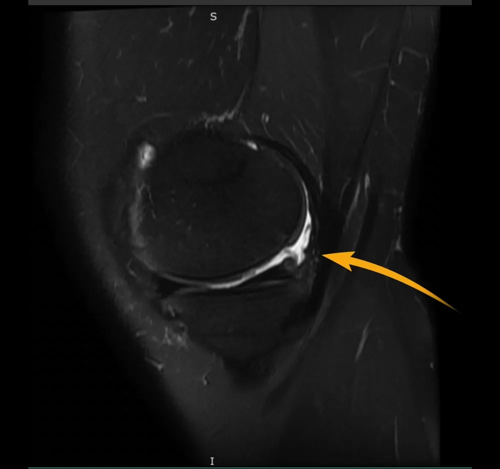
Coronal MRI localized the defect to the medial femoral condyle and confirmed that the rest of the compartment was preserved; the sagittal view showed the lesion’s posterior weight-bearing location and near full-thickness cartilage loss, and the axial view confirmed a posterior central position without marrow edema. After alignment, stability, and meniscal function were reviewed and the treatment options were discussed, the patient elected a single-stage cartilage restoration.
How Dr. Burnham Performs a Two-Implant Agili-C Restoration
The steps shown in the video are:
- Diagnostic arthroscopy. The whole joint is evaluated and any loose bodies, synovitis, or unstable cartilage are addressed. In this case the medial and lateral menisci, including their roots, and both cruciate ligaments were intact, and a focal 12 by 20 millimeter grade 4 lesion was confirmed on the medial femoral condyle.
- Exposure. A limited medial incision and medial parapatellar mini-arthrotomy expose the condyle with the knee in hyperflexion. Direct inspection confirms a well-contained lesion with adequate surrounding bone.
- Planning two sites. Because the lesion is elongated, two 7.5 millimeter sockets are planned to cover it. Both implant centers are marked before any drilling so the preparations do not converge and a bone bridge of at least 5 millimeters is preserved between them.
- Posterior site first. The more difficult posterior position is prepared first. With the alignment guide held perpendicular to the articular surface, a guide pin sets the working axis, the drill sleeve is seated against the surface, and the drill is advanced to its mechanical stop.
- Coaxial reaming and shaping. The reamer is turned clockwise by hand along the same axis, without toggling, until its depth indicator disappears circumferentially. After irrigation, the shaper is advanced along the same axis until its indicator line disappears and it rotates freely at full depth. Reaming, irrigation, and shaping are repeated until the shaper passes without resistance, then the pin is removed and the bed is irrigated again.
- Oblique cartilage margin. The cartilage edge is contoured into a smooth, gently oblique transition rather than a vertical wall, using a cartilage cutter, a number 15 blade, or both. This removes unstable cartilage and prevents the edge from folding into the socket during insertion.
- Insertion by hand. The porous biphasic scaffold has a fixed 10 millimeter depth; the 7.5 millimeter diameter was chosen for this case. With clean gloves the scaffold is oriented and pressed in with gentle force until flush, then a tamper advances it to final depth. No mallet is used. The implant is inspected for fracture, trapped tissue, or cartilage invagination, and a recess of about 2 millimeters below the surrounding surface is confirmed.
- Anterior site. The center and trajectory of the second socket are checked independently before drilling, with the posterior scaffold protected. The same drill, ream, irrigate, and shape sequence is repeated, the margin is refined, and the second scaffold is inserted by hand and seated with the tamper.
- Final inspection and closure. Both implants are confirmed intact, stable, and recessed about 2 millimeters, with an adequate bone bridge and no soft tissue entrapment. The arthrotomy and fascia are closed with 0 Vicryl, the subcutaneous layer with 2-0 Vicryl, and the skin with nylon.

Two details carry most of the technical risk. The first is axis control: every instrument from the guide pin to the shaper must travel the same perpendicular, coaxial path, because a socket that wanders will not hold the scaffold flush and may breach the wall of the adjacent socket. The second is the edge: a vertical cartilage wall tends to fold inward as the scaffold passes, and a trapped flap of cartilage prevents full seating. Saucerizing the margin before insertion solves both problems in advance.
Recovery and Early Follow-Up
In this case, weight bearing was protected for the first 2 to 3 weeks and then advanced progressively, with the goal of full weight bearing by about week 6. Range of motion was allowed as tolerated from the start, physical therapy began on postoperative day 2 or 3, aspirin was prescribed for venous thromboembolism prophylaxis, and the first follow-up visit was at 10 to 14 days. There were no postoperative complications.
At the 2-week visit, AP and lateral radiographs showed maintained joint spaces without fracture or dislocation, with two radiodense scaffold sites visible in the medial femoral condyle in their expected positions and the anticipated anterior soft tissue swelling without a significant effusion. Early radiographs document scaffold position and exclude an acute bony complication. They do not assess biologic integration or cartilage restoration, which is evaluated on MRI at later intervals, as it was in the trial at 12 and 24 months.
Limitations and Open Questions
Agili-C is not a universal cartilage solution. It requires contained lesions with healthy surrounding bone, a perpendicular approach through an arthrotomy, and a disciplined rehabilitation program. The randomized evidence compares it with debridement and microfracture, not with osteochondral allograft or cell-based techniques, and five years is the longest trial follow-up available. Alignment and meniscal status still have to be corrected when they are abnormal; the scaffold cannot compensate for an overloaded compartment. For patients in Baton Rouge and across Louisiana considering cartilage surgery, the honest framing is that this is a well-studied, single-stage option for a defined group of lesions, with results that have held up for five years in a randomized trial and with longer-term data still accumulating.
About the Author
Jeremy M. Burnham, MD is a board-certified orthopedic surgeon and Director of Sports Medicine at Ochsner-Andrews Sports Medicine Institute in Baton Rouge, Louisiana, with a clinical focus on ACL reconstruction, cartilage restoration, and complex knee preservation. Following his orthopedic surgery residency at the University of Kentucky, he completed his sports medicine fellowship at the University of Pittsburgh Medical Center (UPMC), where he trained under the late Dr. Freddie Fu, a pioneer of anatomic ACL reconstruction, Dr. James Bradley, a renowned sports medicine surgeon and longtime professional team orthopedist, and Dr. Volker Musahl, an internationally recognized ACL surgeon and researcher. His team physician experience spans professional sports teams, the University of Pittsburgh, and Southern University. With more than 145 peer-reviewed publications, book chapters, and scientific presentations, Dr. Burnham is the most published ACL surgeon in Louisiana. His research focuses on advancing ACL reconstruction, optimizing return-to-sport outcomes, and pioneering injury prevention, and has been recognized with the Game Changer Award from the Arthritis Foundation and the Playmaker Award from AOSSM. He serves as a site principal investigator for two federally funded clinical trials (NIH STABILITY 2 and Department of Defense STaR Trial). View full credentials and publications.
The Bottom Line
For a contained, high-grade cartilage defect of the femoral condyle or trochlea in a patient with acceptable alignment, stable ligaments, and functioning menisci, the Agili-C scaffold offers a single-stage, off-the-shelf restoration supported by a multicenter randomized trial with five-year follow-up: roughly twice the KOOS improvement of debridement or microfracture at two years, a 77.8% versus 33.6% responder rate, and results that persisted to five years with lower failure rates. It is not a replacement for osteotomy when alignment is the problem, for allograft when bone loss is deep, or for nonoperative care when symptoms have not yet been given a fair trial of it.
When two implants are needed for one elongated lesion, the technique decides the outcome: plan both centers before drilling, keep at least 5 millimeters of bone between them, hold a perpendicular coaxial path through every step, saucerize the cartilage edge, insert by hand, and finish with both scaffolds about 2 millimeters recessed. Patients weighing cartilage surgery can read more about choosing a knee specialist and the questions worth asking before any procedure.
References
- Altschuler N, Zaslav KR, Di Matteo B, et al. Aragonite-Based Scaffold Versus Microfracture and Debridement for the Treatment of Knee Chondral and Osteochondral Lesions: Results of a Multicenter Randomized Controlled Trial. Am J Sports Med. 2023;51(4):957-967. PubMed
- Altschuler N, Zaslav KR, Di Matteo B, et al. Five-Year Follow-up of a Multicenter Randomized Controlled Trial Comparing an Aragonite-Based Scaffold With Microfracture and Debridement for Chondral and Osteochondral Knee Lesions. Am J Sports Med. 2026;54(6):1376-1385. PubMed
- Kon E, Di Matteo B, Verdonk P, et al. Aragonite-Based Scaffold for the Treatment of Joint Surface Lesions in Mild to Moderate Osteoarthritic Knees: Results of a 2-Year Multicenter Prospective Study. Am J Sports Med. 2021;49(3):588-598. PubMed
- Kon E, De Caro F, Dasa V, et al. Female patients report comparable results to males after the implantation of an aragonite-based scaffold for the treatment of knee chondral and osteochondral defects: a gender-based analysis of a RCT at 4 years’ follow-up. J Orthop Traumatol. 2025;26(1):17. PubMed
- Mithoefer K, McAdams T, Williams RJ, Kreuz PC, Mandelbaum BR. Clinical efficacy of the microfracture technique for articular cartilage repair in the knee: an evidence-based systematic analysis. Am J Sports Med. 2009;37(10):2053-2063. PubMed
- Drobnic M, Kon E, Robinson D, Kolar M, Altschuler N. Osteochondral knee joint lesion treated with an aragonite-based scaffold. 12-year follow-up of the world’s first implantation: Case report. J ISAKOS. 2025;16:101040. PubMed
- U.S. Food and Drug Administration. Agili-C Premarket Approval P210034, Summary of Safety and Effectiveness Data. Approved March 29, 2022. Accessed November 8, 2026.
Frequently Asked Questions
What is the Agili-C implant made of, and does it stay in the knee permanently?
Agili-C is a porous scaffold made of aragonite, a natural form of calcium carbonate derived from coral. It has no cells and no metal. Its lower portion sits in bone and its upper portion sits at the cartilage surface. Bone and repair tissue grow into the pores, and the scaffold itself gradually resorbs as the repair matures, so it is not a permanent implant in the way a metal resurfacing device is.
Who is a good candidate for Agili-C cartilage restoration?
The implant is approved for patients 21 to 75 years old with a grade 3 or higher cartilage lesion on the femoral condyle or trochlea totaling 1 to 7 square centimeters, in a knee without severe arthritis. In Dr. Burnham’s practice the lesion must also be well contained, with healthy bone at least 2 millimeters thick around each implant site and no bone cyst deeper than 8 millimeters, and the knee needs acceptable alignment, stable ligaments, and functioning menisci. Patients should have tried nonoperative treatment first.
How does Agili-C compare with microfracture?
In a randomized trial of 251 patients, the scaffold produced about twice the improvement in KOOS score of debridement or microfracture at two years, with 77.8% of implant patients reaching a meaningful improvement versus 33.6% of controls, and a failure rate of 7.2% versus 21.4%. At five years the implant group still scored higher and had fewer failures. The trial did not compare the scaffold with osteochondral allograft or cell-based cartilage procedures, so it does not show superiority over those options.
What is recovery like after a two-implant Agili-C procedure?
Weight bearing is protected for the first 2 to 3 weeks and then advanced progressively toward full weight bearing by about week 6. Knee motion is allowed as tolerated from the start and physical therapy begins within a few days of surgery. A blood thinner such as aspirin is typically used for clot prevention, and the first follow-up visit is at 10 to 14 days. Return to higher-impact activity depends on healing seen on later imaging and on strength and function in rehabilitation, and is individualized.
Why were two implants used instead of one?
The lesion in this case measured 12 by 20 millimeters, longer than a single 7.5 millimeter scaffold could cover. Two scaffolds placed in line covered the elongated defect while leaving a bridge of healthy bone at least 5 millimeters wide between them. Planning both implant centers before drilling keeps the two sockets from converging and preserves the bone that supports each implant.