
A sudden twist on the basketball court, an awkward landing from a jump, a sharp change of direction on the soccer field, and then it happens: a loud “pop” in the knee, followed by immediate swelling and the unsettling feeling that the joint cannot be trusted. These are the hallmark signs of an anterior cruciate ligament (ACL) tear, one of the most common and consequential knee injuries in sports medicine. Approximately 200,000 ACL injuries occur each year in the United States, affecting athletes, active adults, and weekend warriors alike.
Recognizing ACL tear symptoms early can make a meaningful difference in treatment outcomes. Research from the ACL reconstruction literature, including a multicenter study by the PIVOT Study Group with Dr. Burnham as a co-investigator (Lian, Burnham et al., JBJS 2020), has shown that the degree of knee instability exists on a continuum, and understanding where an injury falls on that spectrum is critical for determining the best path forward.
In This Article
What Is the ACL and Why Does It Matter?
The anterior cruciate ligament is a band of dense connective tissue that runs diagonally through the center of the knee, connecting the thighbone (femur) to the shinbone (tibia). Its primary job is to prevent the tibia from sliding forward under the femur and, critically, to control rotational stability of the knee. The ACL consists of two functional bundles (the anteromedial bundle and the posterolateral bundle), each contributing to stability at different angles of knee flexion.
When the ACL tears, the knee loses its primary restraint against rotational forces. As Dr. Burnham’s research with the PIVOT Study Group has demonstrated, the resulting instability is measurable and progressive: partial tears produce less rotational laxity than complete tears, which in turn produce less than failed reconstructions (Lian, Burnham et al., 2020). This progressive instability is why early and accurate diagnosis matters: the longer an unstable knee goes unrecognized, the greater the risk of secondary damage to the meniscus and cartilage.
The 5 Most Common ACL Tear Symptoms
1. A “Pop” at the Moment of Injury
The most widely recognized ACL tear symptom is an audible or felt “pop” in the knee at the time of injury. Patients frequently describe it as a loud snap that other people nearby can hear. The pop corresponds to the sudden failure of the ligament fibers under mechanical stress, and it is reported in roughly 50 to 70 percent of confirmed ACL tears. While not every ACL tear produces a pop (and not every knee pop indicates an ACL tear), this symptom, combined with the ones below, is highly suggestive of ligament injury.
2. Rapid Knee Swelling Within Hours
An ACL tear disrupts blood vessels within the ligament, causing blood to accumulate inside the joint (hemarthrosis). This produces noticeable swelling that typically develops within the first two to four hours after injury. The speed of onset is a distinguishing feature: a simple sprain or bruise may take a day or more to swell, while an ACL tear causes the knee to balloon rapidly. Clinical studies have shown that a tense hemarthrosis within six hours of a knee injury has a 70 to 80 percent chance of being associated with an ACL tear.
This swelling is technically called a knee effusion, meaning fluid has accumulated inside the joint capsule itself rather than in the soft tissues around the knee. When an orthopedic surgeon examines an acutely injured knee, one of the first things assessed is whether an effusion is present. A “ballotable patella” (the kneecap can be pushed down and bounces back up) or a positive “fluid wave test” confirms that the swelling is intra-articular. The presence of a large, tense effusion after a twisting injury is one of the strongest clinical indicators that a significant internal knee injury, most commonly an ACL tear, has occurred. In many cases, the surgeon may aspirate (drain) the effusion in the office to relieve pressure, improve comfort, and allow a more accurate physical examination of ligament stability.
3. Knee Instability or “Giving Way”
After the initial pain and swelling begin to subside (often within one to two weeks), many patients notice that the knee feels unstable, particularly during cutting, pivoting, or decelerating movements. The knee may buckle or “give way” without warning. This instability reflects the loss of the ACL’s rotational control. Research by Dr. Burnham and colleagues has quantified this phenomenon using validated measurement tools, showing that side-to-side differences in lateral compartment translation averaged 2.5 mm in patients with complete ACL tears, compared to 1.4 mm in partial tears (Lian, Burnham et al., 2020). The sensation of giving way is the single strongest clinical indicator that the ACL may be compromised.
4. Pain, Especially with Weight-Bearing and Pivoting
ACL tear pain is often sharp and intense at the moment of injury, then gradually transitions to a deep, aching discomfort over the following days. The pain is typically diffuse across the knee rather than pinpointed to one spot. Interestingly, many patients report that the pain actually decreases after the first few days as the acute inflammatory response settles, which can create a false sense of recovery. Pain tends to return when the patient attempts to resume activity, particularly movements that stress the ACL like twisting, cutting, or decelerating from a run.
5. Loss of Range of Motion
Swelling and pain combine to restrict the knee’s ability to fully bend or straighten after an ACL tear. Patients often notice difficulty achieving full extension (straightening the knee completely) due to the hemarthrosis and guarding. If a torn meniscus accompanies the ACL tear (which occurs in approximately 40 to 60 percent of cases), a “mechanical block” may prevent full bending or straightening. Patients with combined ACL and meniscus injuries often present with more pronounced motion loss and may notice catching or locking of the knee joint.
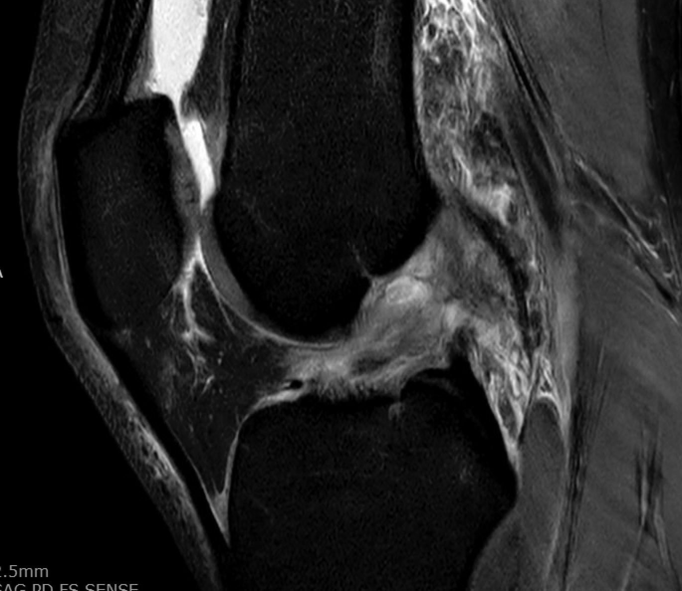
Partial ACL Tear vs. Complete ACL Tear
Not all ACL injuries are the same. The ACL can sustain a partial tear (involving one of its two bundles while the other remains intact) or a complete tear (both bundles disrupted). Between 10 and 28 percent of ACL injuries are estimated to be partial tears. Patients with a partial ACL tear often present with milder symptoms: the pop may be less dramatic, swelling may be moderate rather than severe, and the knee may feel more stable during daily activities. However, the distinction matters for treatment planning.
The PIVOT Study Group’s multicenter analysis of 304 patients provided quantitative evidence for this continuum. Using validated measurement tools to assess rotational laxity under anesthesia, the investigators found a stepwise increase in knee instability: partial ACL tears showed a side-to-side difference of 1.4 mm in lateral compartment translation, complete tears showed 2.5 mm, and failed ACL reconstructions showed 3.3 mm (p = 0.01) (Lian, Burnham et al., 2020). Importantly, up to 42 percent of partial tears may progress to complete tears over time, even after seemingly minor twisting events, which is why a partial tear still warrants careful evaluation and monitoring.

How an ACL Tear Is Diagnosed
Physical Examination: The Lachman, Pivot Shift, and Lever Sign Tests
An experienced orthopedic surgeon can often diagnose an ACL tear based on clinical examination alone. Three physical tests form the standard diagnostic toolkit:
The Lachman test is performed with the knee bent to approximately 20 to 30 degrees. The examiner stabilizes the thigh with one hand and pulls the lower leg forward with the other. In an ACL-deficient knee, the tibia translates forward with a soft or absent endpoint. Meta-analyses have reported the Lachman test’s sensitivity at 81 to 87 percent and specificity at 85 to 94 percent (Sokal et al., 2022; Huang et al., 2016).
The pivot shift test reproduces the rotational instability that patients experience as “giving way.” The examiner applies internal rotation and a valgus force to the extended knee, then flexes it. A positive pivot shift produces a palpable clunk as the tibia reduces. The pivot shift is the most specific test for ACL tears, with a pooled specificity of 94 to 98 percent, though its sensitivity is lower at 24 to 55 percent because muscle guarding in an awake patient can mask the shift (Benjaminse et al., 2006; Sokal et al., 2022). This is why the pivot shift is most reliably assessed under anesthesia during surgery.
The lever sign test is a newer addition to the diagnostic toolkit. With the patient lying flat, a fist is placed under the proximal calf to act as a fulcrum. In an intact knee, the heel lifts off the table; in an ACL-deficient knee, the heel stays down. Recent meta-analysis data show the lever sign has a sensitivity of 83 percent and specificity of 91 percent (Sokal et al., 2022).
MRI: Confirming the Diagnosis
Magnetic resonance imaging (MRI) is the gold standard imaging study for confirming an ACL tear and evaluating associated injuries. MRI can visualize the ligament directly and detect tears with a sensitivity of approximately 94 to 95 percent and specificity of 94 to 95 percent. Beyond confirming the ACL tear itself, MRI reveals critical associated findings that influence treatment planning: meniscal tears, cartilage damage, bone bruise patterns, and the condition of other ligaments.
Bone bruise patterns on MRI are particularly informative. The classic ACL tear bone bruise pattern involves the lateral femoral condyle and the posterolateral tibial plateau, reflecting the pivot shift mechanism of injury. Dr. Burnham’s research on bony morphologic factors has highlighted how specific anatomic features visible on MRI and radiographs, including posterior tibial slope and intercondylar notch width, influence both ACL injury risk and rotational stability after reconstruction (Burnham et al., 2017).
When to See an Orthopedic Surgeon
Any combination of a pop, rapid swelling, and knee instability after an injury warrants prompt evaluation by an orthopedic surgeon with sports medicine expertise. The urgency increases when the knee gives way during weight-bearing, when swelling does not improve within several days, when the patient is an athlete who needs to return to cutting and pivoting sports, or when the patient is a young athlete whose growth plates are still open.
Delaying evaluation carries real consequences. Each episode of giving way can cause additional damage to the meniscus and articular cartilage. Studies have consistently shown that patients who undergo ACL reconstruction more than six months after injury have a significantly higher rate of meniscal tears compared to those treated earlier. Understanding the cost of ACL reconstruction surgery can also help patients plan their treatment timeline and financial considerations. At Ochsner-Andrews Sports Medicine Institute in Baton Rouge, Dr. Burnham and the sports medicine team use the latest evidence and advanced diagnostic tools, including quantitative pivot shift measurement, to tailor each patient’s treatment plan to their specific injury pattern, anatomy, and activity goals.
About the Author: Dr. Jeremy Burnham is a board-certified orthopedic surgeon and sports medicine specialist at Ochsner-Andrews Sports Medicine Institute in Baton Rouge, Louisiana. With 53 peer-reviewed publications including multicenter research on ACL injury diagnosis and treatment through the PIVOT Study Group, Dr. Burnham brings fellowship-trained expertise from the University of Pittsburgh Medical Center to patients throughout Louisiana and the Gulf South.
The Bottom Line
The combination of a pop at the time of injury, rapid knee swelling, and a feeling of instability is the classic presentation of an ACL tear. Partial tears and complete tears exist on a measurable continuum of instability, and distinguishing between them requires expert clinical examination supplemented by MRI. An experienced orthopedic surgeon can diagnose most ACL tears in the office using the Lachman, pivot shift, and lever sign tests. Early evaluation is important because ongoing instability can lead to additional meniscal and cartilage damage that complicates future treatment. If these symptoms sound familiar after a recent knee injury, scheduling an evaluation with a sports medicine specialist is the most important next step. Once an ACL tear is confirmed, you can compare your ACL graft options with your surgeon to determine the best reconstruction approach for your specific injury and activity level. Learning how to choose an ACL surgeon will help ensure you receive expert care tailored to your needs.
References
- Lian J, Diermeier T, Meghpara M, Popchak A, Smith CN, Kuroda R, Zaffagnini S, Samuelsson K, Karlsson J, Irrgang JJ, Musahl V; PIVOT Study Group. Rotatory Knee Laxity Exists on a Continuum in Anterior Cruciate Ligament Injury. J Bone Joint Surg Am. 2020;102(3):213-220. PMID: 31876642.
- Sokal PA, Norris R, Maddox TW, Oldershaw RA. The diagnostic accuracy of clinical tests for anterior cruciate ligament tears are comparable but the Lachman test has been previously overestimated: a systematic review and meta-analysis. Knee Surg Sports Traumatol Arthrosc. 2022;30(10):3287-3303. PMID: 35150292.
- Huang W, Zhang Y, Yao Z, Ma L. Clinical examination of anterior cruciate ligament rupture: a systematic review and meta-analysis. Acta Orthop Traumatol Turc. 2016;50(1):22-31. PMID: 26854045.
- Benjaminse A, Gokeler A, van der Schans CP. Clinical diagnosis of an anterior cruciate ligament rupture: a meta-analysis. J Orthop Sports Phys Ther. 2006;36(5):267-288. PMID: 16715828.
- Burnham JM, Pfeiffer T, Shin JJ, Herbst E, Fu FH. Bony morphologic factors affecting injury risk, rotatory stability, outcomes, and re-tear rate after anterior cruciate ligament reconstruction. Ann Joint. 2017;2:44.
Frequently Asked Questions
What does a torn ACL feel like?
Most patients describe an ACL tear as a sudden pop or snap in the knee during a pivoting, cutting, or landing movement. Immediate pain is common but often subsides within a few days. The knee typically swells within two to four hours. Over the following weeks, the most telling symptom is instability: the feeling that the knee buckles or gives way during activities that require twisting or quick direction changes.
Can you still walk with a torn ACL?
Many patients can walk on a torn ACL once the initial pain and swelling improve, which typically takes one to two weeks. u003ca href=u0022/can-you-walk-on-torn-acl/u0022u003eWalkingu003c/au003e in a straight line does not heavily stress the ACL because forward motion is relatively stable. However, activities that involve pivoting, cutting, going up or down stairs, or sudden stops often provoke the instability and giving way that characterize an ACL-deficient knee. Being able to walk does not mean the ACL is intact.
How soon should I see a doctor after a suspected ACL tear?
Evaluation within the first one to two weeks of injury is recommended. While an ACL tear is not a medical emergency, early diagnosis allows for appropriate bracing, initiation of pre-surgical rehabilitation (which improves surgical outcomes), and protection of the meniscus and cartilage from further damage caused by ongoing instability episodes. Research has shown that delaying ACL reconstruction beyond six months increases the rate of associated meniscal tears.
What is the difference between an ACL sprain and an ACL tear?
The terms overlap significantly. An ACL u0022sprainu0022 technically refers to any stretch or damage to the ligament, graded on a scale from Grade I (mild stretch, fibers intact) to Grade II (partial tear, some fibers disrupted) to Grade III (complete tear). In practice, when patients or physicians refer to a u0022torn ACL,u0022 they typically mean a Grade III (complete) injury. A Grade II injury is what surgeons call a partial ACL tear. Both Grade II and Grade III injuries can cause instability and may require surgical treatment depending on the patient’s activity level and goals.
Do all ACL tears require surgery?
Not all ACL tears require surgery, but the decision depends on several factors: the patient’s age, activity level, degree of instability, associated injuries (such as meniscal tears), and personal goals. Young, active patients who want to return to cutting and pivoting sports generally benefit from ACL reconstruction. Older or less active patients who are willing to modify their activities may manage well with rehabilitation alone. An orthopedic surgeon with sports medicine expertise can help weigh these factors for each individual case.