In the world of sports medicine, innovation and precision are key to helping athletes return to their peak performance. One such advancement is the use of amniotic membrane tissue during ACL reconstruction, a technique Dr. Jeremy Burnham has pioneered to address the challenge of quadriceps muscle weakness post-surgery. This video explores the rationale and surgical technique behind applying amniotic grafts to the quad tendon harvest site.

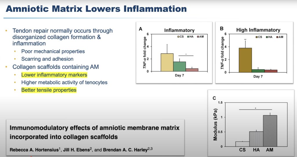
Amniotic membrane, derived from donated umbilical cord and placenta tissue after live births (with no harm to babies), is rich in growth factors like TIMP2 and TIMP3, which are critical for tendon repair and reducing inflammation. Drawing inspiration from its successful use in foot and ankle surgery—particularly for Achilles tendon procedures—Dr. Burnham, a board-certified orthopedic surgeon and sports medicine specialist at Ochsner-Andrews Sports Medicine Institute, adapted this approach for ACL reconstruction using quadriceps tendon autografts. Laboratory and clinical studies have shown that amniotic matrix enhances tenocyte proliferation, improves tendon healing, and supports nerve healing and muscle growth, making it a promising tool for optimizing recovery.

The quadriceps tendon is increasingly favored for ACL reconstruction due to its reliable sizing and superior collagen density. However, a common hurdle, known as arthrogenic muscle inhibition, can lead to lingering quad weakness. Dr. Burnham’s technique, inspired by the agricultural concept of cover cropping—replenishing nutrients after a harvest—applies an amniotic graft to the quad tendon harvest site to restore vital growth factors and promote healing. “We’re mimicking nature,” Dr. Burnham notes, echoing the philosophy of his mentor, Dr. Freddy Fu, to enhance the body’s natural recovery processes.
In the surgical video, Dr. Burnham demonstrates this meticulous process. Beginning with a 2cm incision proximal to the patella, the quadriceps tendon is exposed, and a partial-thickness graft is carefully harvested using a double-blade knife and specialized tools like a number nine scalpel. The defect is closed with absorbable sutures, and a 3×8 cm Arthrex amniotic graft is prepared and oriented correctly. The graft is then shuttled into the harvest site, secured with sutures and an arthroscopic knot, ensuring it integrates seamlessly to support tendon regeneration. The procedure concludes with standard ACL reconstruction, setting the stage for robust recovery.
By 16 weeks post-surgery, this patient exhibits impressive quadriceps symmetry. Between three to four months, they progress to functional exercises like squats, plyometric jumps, and running, with some, like the cheerleader highlighted in the video, returning to sport-specific activities by four to five months. This innovative approach underscores the power of combining advanced surgical techniques with a patient-centered focus on recovery.
#SportsMedicine #ACLRecovery #OrthopedicInnovation #AthleteRecovery #BatonRougeAthletes
